Difference between revisions of "Multimodal microscopy"
Jump to navigation
Jump to search
| Line 6: | Line 6: | ||
<gallery widths=200px heights=150px> | <gallery widths=200px heights=150px> | ||
| − | + | File:microlentille.jpg|3D profil of a Fresnel micro-lens | |
| − | + | File:microscopie_4D.jpg|3D Profil of a Hall micro-sensor | |
| − | + | File:BaslerALT3Dv4c.gif|Real time aquired 3D image | |
</gallery> | </gallery> | ||
Revision as of 16:35, 5 October 2014
The interferometric microscopy is an historical subject of the team.
What is multimodal microscopy?

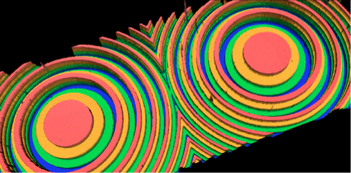
3D profil of a Fresnel micro-lens

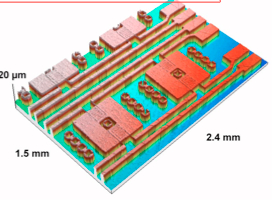
3D Profil of a Hall micro-sensor

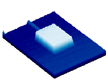
Real time aquired 3D image
- Scanning interference microscopy is a powerful technique based on far field imaging and interferometry that can be used for extracting information on micro- and nanostructures embedded in complex materials, devices and microsystems. Two topics are covered: 4D microscopy and techniques for complex layer analysis.
- 4D microscopy (3D+time) enables the 3D measurement in real time of surfaces that evolve over time, such as those found in soft materials, MicroElectroMechanical Systems (MEMS) and chemical reactions.
- The second topic addresses the challenges of using interferometry to measure thick, semi-transparent or translucent layers of materials such as hydroxyapatite (biomaterials), colloids and polymers. These techniques are also useful for characterising nanostructured photonic devices developed in the theme Photonics Modeling and Simulation.