Difference between revisions of "Multimodal microscopy"
Montgomery (talk | contribs) |
|||
| Line 1: | Line 1: | ||
| − | '''P. Montgomery, M. Flury''', F. Anstotz, S. Lecler, V. Maioli, D. Montaner, F. Salzenstein. | + | '''P. Montgomery, M. Flury''', F. Anstotz, S. Lecler, V. Maioli, D. Montaner, A. Nahas, F. Salzenstein. |
| − | |||
| − | '''What is multimodal | + | '''What is multimodal nanoscopy?''' |
| + | |||
| + | The IPP team has been at the forefront of research in the field of interference microscopy for many years. The aims are several. More recently, they concern improving the lateral resolution to beyond the diffraction limit without using markers. The addition of different imaging modalities allows using the same imaging system to access different types of information such as the microscopic surface topography (static and moving), the high-resolution structure of transparent layers and the local spectroscopic response. The use of dedicated environmental chambers allows the study of specific parameters of samples in a controlled environment. The study of specific algorithms allows optimisation of fringe processing and the extraction of the required information. | ||
| + | |||
| + | The team is also extending its field of investigation of samples from the historical application areas of materials and micro- nanotechnologies to those of the biological field. It does this through strong links with local actors in the field (ICS, IPCMS, IGBMC, IPHC, etc.), starting from the fundamental understanding of the basic principles involved, through the development of prototype instrumentation, right through to the technology transfer of the techniques developed. | ||
<gallery widths=200px heights=150px> | <gallery widths=200px heights=150px> | ||
| Line 13: | Line 16: | ||
</gallery> | </gallery> | ||
| − | :Scanning interference microscopy is a powerful technique based on far field | + | :Scanning interference microscopy is a powerful technique based on far field optical reflection microscopy and interferometry that can be used for extracting information on micro- and nanostructures embedded in complex materials, devices and microsystems. While the technique is classically used for measuring static microscopic surface roughness and topography, we have developed other measurement modes and various techniques for improving the measurements. |
| + | :The following topics are covered: | ||
:4D microscopy (3D+time) enables the 3D measurement in real time of surfaces that evolve over time, such as those found in soft materials, MicroElectroMechanical Systems (MEMS) and chemical reactions. | :4D microscopy (3D+time) enables the 3D measurement in real time of surfaces that evolve over time, such as those found in soft materials, MicroElectroMechanical Systems (MEMS) and chemical reactions. | ||
Revision as of 16:48, 2 August 2022
P. Montgomery, M. Flury, F. Anstotz, S. Lecler, V. Maioli, D. Montaner, A. Nahas, F. Salzenstein.
What is multimodal nanoscopy?
The IPP team has been at the forefront of research in the field of interference microscopy for many years. The aims are several. More recently, they concern improving the lateral resolution to beyond the diffraction limit without using markers. The addition of different imaging modalities allows using the same imaging system to access different types of information such as the microscopic surface topography (static and moving), the high-resolution structure of transparent layers and the local spectroscopic response. The use of dedicated environmental chambers allows the study of specific parameters of samples in a controlled environment. The study of specific algorithms allows optimisation of fringe processing and the extraction of the required information.
The team is also extending its field of investigation of samples from the historical application areas of materials and micro- nanotechnologies to those of the biological field. It does this through strong links with local actors in the field (ICS, IPCMS, IGBMC, IPHC, etc.), starting from the fundamental understanding of the basic principles involved, through the development of prototype instrumentation, right through to the technology transfer of the techniques developed.

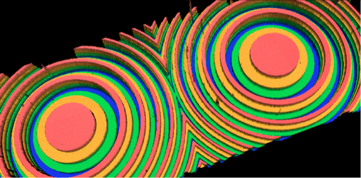
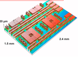
3D profil of a Fresnel micro-lens

3D Profil of a Hall micro-sensor

Real-time aquisition of a 3D image
- Scanning interference microscopy is a powerful technique based on far field optical reflection microscopy and interferometry that can be used for extracting information on micro- and nanostructures embedded in complex materials, devices and microsystems. While the technique is classically used for measuring static microscopic surface roughness and topography, we have developed other measurement modes and various techniques for improving the measurements.
- The following topics are covered:
- 4D microscopy (3D+time) enables the 3D measurement in real time of surfaces that evolve over time, such as those found in soft materials, MicroElectroMechanical Systems (MEMS) and chemical reactions.
- The second topic addresses the challenges of using interferometry to measure thick, semi-transparent or translucent layers of materials such as hydroxyapatite (biomaterials), colloids and polymers. These techniques are also useful for characterising nanostructured photonic devices developed in the theme Photonics Modeling and Simulation.
Funding:
- 2 Scholarships of the Indonesian government
- EOTECH SA
- ICube internal call
- CAM4D Project (SATT Conectus Alsace)
Past:
- PICS (CNRS)
- Coopération (CNRS)
- EU INTERREG III
- ACO (CNRS) in collaboration with ESPCI ParisTech (Paris)